How Indian scientists are mapping the brain’s last frontier

How Indian Scientists Are Mapping the Brain’s Last Frontier
Ninoda.com – How Indian scientists are mapping the brain has become a pivotal endeavor in the field of neuroscience, addressing one of the most complex challenges in modern science. With the human brain containing an estimated 86 billion neurons, understanding its intricate structure and function remains a daunting task. For over a century, researchers have approached this challenge by combining macroscopic imaging techniques with microscopic analysis, akin to early explorers charting uncharted lands. However, the lack of seamless integration between these methods has left critical gaps in our comprehension of the brain’s last frontier—the brainstem. Despite advances in imaging technologies, medical professionals diagnosing conditions like Alzheimer’s often rely on limited tissue samples, which fail to capture the full complexity of neural networks. How Indian scientists are mapping the brain now aims to change this by creating a more detailed and accessible framework for studying this vital region.
A New Breakthrough in Brain Mapping
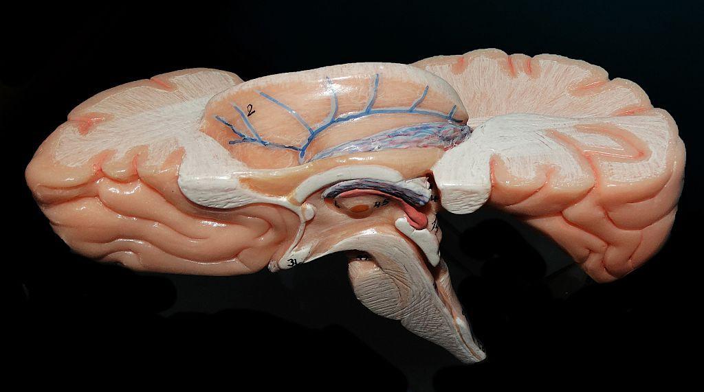
Indian scientists at the Sudha Gopalakrishnan Brain Centre (SGBC), affiliated with the Indian Institute of Technology, Madras (IIT-M), have made a groundbreaking achievement in the field of brain mapping. Their recent development of the world’s most comprehensive three-dimensional atlas of the human brainstem has set a new standard for neuroscientific research. This digital map provides an unprecedented level of detail, allowing researchers to visualize the brainstem at the cellular level. By merging high-resolution microscope images with neurochemical data, the atlas bridges the gap between large-scale brain imaging and microscopic examination. The project, known as Anchor, not only offers a cellular-level view of the brainstem but also enables scientists to navigate seamlessly from macroscopic scans to individual nerve cells, revolutionizing how we study brain anatomy.
What makes this breakthrough particularly significant is its integration of data from fetal, childhood, and adult brains, creating a dynamic map that reflects developmental changes. The atlas includes over 500 tissue slices, each meticulously analyzed to identify more than 200 distinct cell clusters and nerve pathways. This level of detail is made possible by the use of advanced 3D reconstruction techniques, which allow for precise alignment of microscopic findings with macroscopic structures. The inclusion of eight chemical markers further differentiates cell types, providing a clearer picture of the brainstem’s role in essential functions such as breathing, heartbeat, and consciousness. How Indian scientists are mapping the brain in this way not only enhances our understanding of its structure but also paves the way for more accurate diagnoses and treatments of neurological disorders.
Technology and Collaboration
How Indian scientists are mapping the brain has been made possible through a combination of cutting-edge technology and cross-disciplinary collaboration. The Anchor project leverages high-resolution imaging and machine learning algorithms to process vast amounts of data efficiently. This technology ensures that each tissue slice is analyzed with precision, capturing the subtle variations in cell structure and function that were previously overlooked. The collaboration between the SGBC and IIT-M has been instrumental in developing this tool, as it combines expertise in neuroscience, bioinformatics, and engineering. Such partnerships highlight the importance of interdisciplinary research in tackling complex scientific problems.
One of the key challenges in brain mapping is maintaining the integrity of cellular structures while integrating data from different sources. The Indian team has overcome this by using advanced imaging techniques that preserve the spatial relationships between cells. This has enabled them to create a comprehensive atlas that serves as a foundational resource for future research. The Anchor project also emphasizes the use of open-access platforms, ensuring that the data is freely available to neuroscientists, neurologists, and surgeons worldwide. This approach not only democratizes access to critical research but also fosters global collaboration in advancing neuroscience. How Indian scientists are mapping the brain in this manner could inspire similar initiatives in other regions, setting a benchmark for precision and accessibility in the field.
“This is a visionary programme that positions India as a leader in neuroscience,” said Shubha Tole, a neuroscientist at the Tata Institute of Fundamental Research. “The work represents an unprecedented integration of engineering, medicine, and brain research, offering a unique perspective on the brainstem’s complexity.” The project’s success underscores the growing role of Indian institutions in contributing to global scientific advancements. By creating a digital map that aligns macroscopic and microscopic views, the team has provided a valuable tool for understanding the brain’s fundamental functions and the disorders that affect them.
Applications in Medical Research
How Indian scientists are mapping the brain has far-reaching implications for medical research and clinical applications. The Anchor atlas could revolutionize the diagnosis and treatment of conditions such as Parkinson’s disease, stroke, and sudden infant death syndrome (SIDS). By providing a detailed view of the brainstem’s cellular architecture, the project offers insights into the mechanisms underlying these disorders, potentially leading to more targeted therapeutic interventions. For instance, understanding the specific nerve pathways involved in breathing and heartbeat regulation could enhance treatments for neurodegenerative diseases that affect these vital functions.
Moreover, the atlas serves as a critical resource for researchers studying the brain’s development and aging processes. Its ability to map the brainstem across different life stages allows for comparative analysis, which is essential in uncovering how neural structures evolve over time. This information can be used to develop better models of brain function and dysfunction, ultimately improving our ability to predict and manage neurological conditions. While the Anchor project is not a diagnostic tool itself, it provides a framework that could be integrated with existing technologies to enhance accuracy and efficiency in medical practice.
Looking ahead, the work of how Indian scientists are mapping the brain is expected to inspire further innovations in neuroscience. The project’s open-access nature encourages other researchers to build upon its findings, fostering a collaborative environment that accelerates discovery. As the atlas becomes a standard reference, it may also lead to the development of new tools and methodologies for studying the brain’s last frontier. The potential for this work to transform our understanding of the human brain is immense, and its impact on both research and medicine is likely to be profound. By combining precision with accessibility, the Anchor project exemplifies how Indian scientists are making significant strides in this vital field.